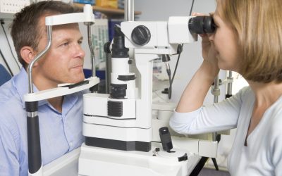
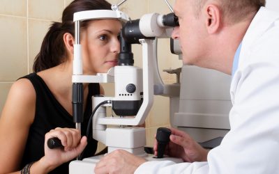
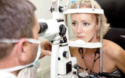
You may have been told that you might want to consider surgery for a visual error or disease. Usually this diagnosis is made during an eye exam with help from a slit lamp microscope. This type of device uses bright light to give the ophthalmologist a closer view of the various structures at the front or inside the eye. It is used as a primary tool for determining eye health and detecting disease.
Preparing for a Slit Lamp Exam
If you have not done so already, you should schedule an eye exam. You do not need to prepare for the assessment when a slit lamp is used to determine whether or not eye surgery may be needed. However, your will be given dilating drops and therefore need to have someone drive you home after this type of test.
When the exam is performed, the eye doctor sits in front on the slit lamp. He or she asks the patient to place his or her chin in the chin rest and position the brow in a forehead band. Placing your head in this manner keeps it steady during the assessment.
What the Doctor Checks
When performing this examination, the doctor looks at the sclera (the white part of the eye), the cornea, and the lens at the front. He or she then looks at the back of the eye, reviewing the retina and optic nerve. As you can see, this type of thorough assessment can indeed indicate any need for eye surgery.
For example, examination of the optic nerve in the back of the eye, can help the doctor assess if a patient has glaucoma. Looking at the retina can assist in the diagnosis of age-related macular degeneration. He or she can also look for corneal abrasions at the front of the eye or see if you are a good candidate for LASIK eye surgery in Orlando, FL.
Where to Get Further Details
Would you like to know more about this type of eye exam process? If so, contact Hunter Vision online for further details today. The more you know about the instruments that are used, the easier it will be to plan for the exam and find out if you are indeed the right candidate for surgery.


